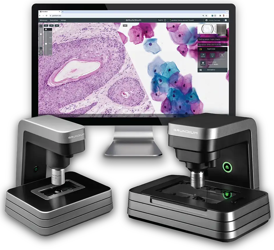


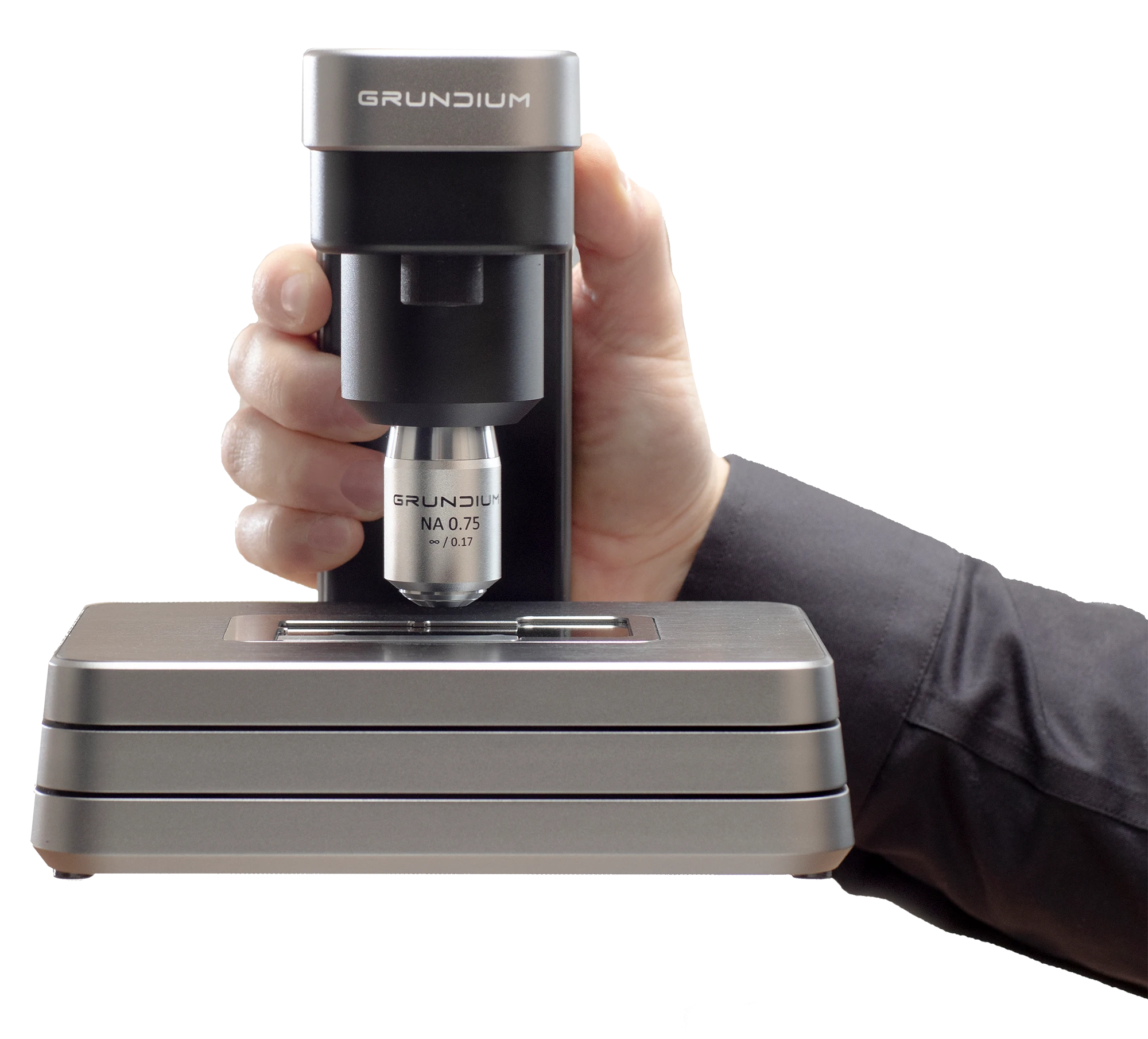
A digital microscope scanner, or digital pathology scanner, converts traditional glass microscope slides into high-resolution digital images that can be viewed, reviewed, and shared remotely. In practice, Grundium’s compact Ocus® scanners, such as the Ocus®20, make digitized slides accessible through a web browser, supporting workflows such as clinical review where permitted, second opinions, research, and education while reducing reliance on physical slide handling and transport.
Whole slide scanners are used to digitize complete microscope slides for case review, documentation, and sharing across teams. They are commonly used in workflows such as histology and cytology review, second opinions, teaching, and research. With the Grundium Ocus® series, digitized slides can be accessed and shared through a web browser, supporting flexible workflows and helping reduce the need to move physical slides between locations.
Digital pathology scanners allow digitized slides to be accessed and shared through a web browser, enabling specialists to review cases without being in the same location. This supports remote consultations, second opinions, and collaboration across laboratories, helping reduce reliance on physical slide transport and supporting faster access to expert input.
Digital pathology imaging can streamline workflows by enabling rapid access to digitized slides, reducing transport-related delays, and supporting remote collaboration. This helps laboratories and research teams improve process efficiency and maintain more consistent workflows.
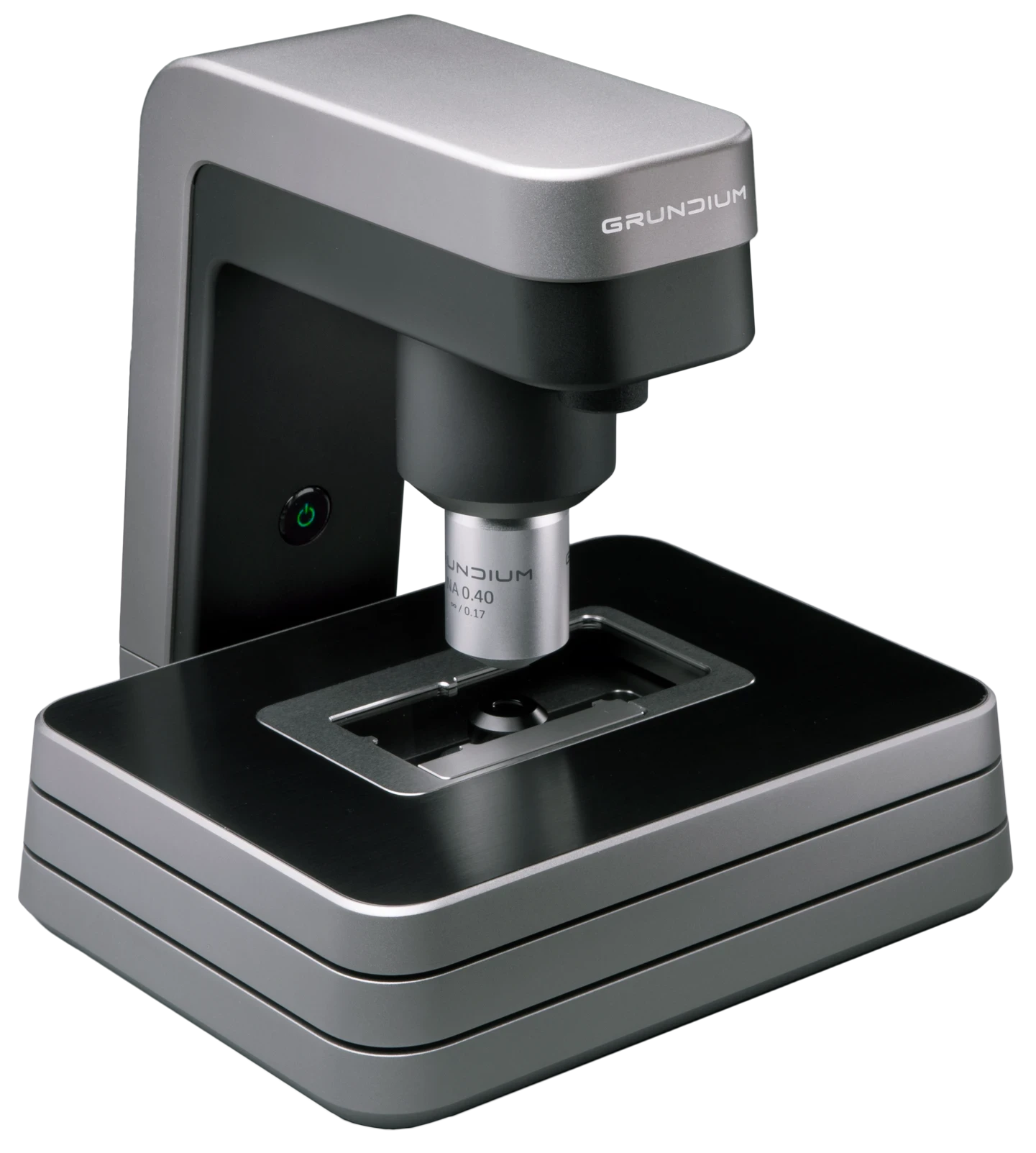
When choosing a digital pathology scanner, important considerations include image quality, magnification, scanning speed, ease of use, workflow compatibility, slide capacity, and integration into daily operations. Grundium’s compact, browser-based Ocus® scanners are designed to deliver high-resolution imaging while keeping setup, training, and infrastructure requirements low.


