9 questions and answers about whole slide imaging

Divider

In the dynamic world of medical diagnostics, Whole Slide Imaging (WSI) emerges as a game-changer in pathology. Many professionals grapple with its nuances as the shift to digital pathology accelerates. Dive into our concise guide, addressing nine pivotal questions about WSI. Equip yourself with insights and stay updated in this digital evolution.
Introduction to whole slide imaging

Whole Slide Imaging, WSI, is a groundbreaking technology that is transforming the field of pathology. By digitizing entire glass slides, WSI allows pathologists to view and analyze high-resolution images on a computer screen, enhancing the accuracy and efficiency of diagnoses. This innovative approach not only facilitates remote consultations and second opinions but also integrates seamlessly with digital pathology workflows, paving the way for advancements in personalized medicine.

The significance of WSI in medical diagnostics cannot be overstated. It offers unprecedented opportunities for education, research, and clinical practice by providing a platform for the storage, sharing, and analysis of digital slides. As pathology departments worldwide adopt this technology, WSI is revolutionizing traditional practices, making pathology more accessible and collaborative than ever before.

As you explore the following nine essential questions about Whole Slide Imaging, consider how this technology is reshaping the landscape of pathology and what it means for the future of medical diagnostics.

1. HOW DOES WHOLE SLIDE IMAGING (WSI) QUALITY COMPARE TO TRADITIONAL MICROSCOPY IN TERMS OF RESOLUTION AND DETAIL?

Whole Slide Imaging and traditional microscopy serve the same purpose of visualizing tissue samples for pathological examination, but there are differences in how they present the visual data.

In terms of resolution, the quality of a WSI image is generally high, comparable to what can be seen under a microscope. Many WSI systems are capable of scanning slides at 20x or 40x magnification, creating images with fine detail. The resolution of these images is typically sufficient for most diagnostic purposes.

However, it is essential to note that some subtleties visible under traditional microscopy may not always be captured as effectively with WSI. For instance, the three-dimensionality of specific cellular structures can be more apparent under a microscope, as the pathologist can manually adjust the focus to visualize different planes of the specimen. On the other hand, WSI scanners typically focus on a single plane. This issue can sometimes be mitigated by using scanners that provide z-stacking capabilities, creating a series of images at different focal planes, but this approach is not always practical due to the resulting increase in file size and processing time.

On the other hand, WSI offers the advantage of easy navigation, allowing pathologists to quickly move to different areas of a slide or toggle between different slides. The images can also be zoomed in or out smoothly, which can provide a different perspective on tissue morphology.

2. WHAT IS THE LEARNING CURVE LIKE FOR PATHOLOGISTS ADAPTING TO WHOLE SLIDE IMAGING?

The transition from traditional microscopy to Whole Slide Imaging does involve a learning curve for pathologists, but the exact steepness of that curve can vary based on factors such as prior experience with digital tools, the complexity of the WSI system, and the level of training and support provided.

Initially, pathologists may need to familiarize themselves with the WSI system, learning how to navigate and manipulate digital slides and use the software's features effectively. They may also need to adapt to interpreting digital images, recognizing digital artifacts, and understanding the limitations of WSI.

Moreover, many find that WSI can enhance their workflows once they've adjusted to the new technology. The ability to easily share slides for consultations, integrate digital pathology data with other health records, and use digital tools for annotations and measurements can be significant advantages.
To ease the learning curve, it's crucial that pathologists receive comprehensive training when transitioning to WSI. This training should ideally include technical instruction on using the system and guidance on interpreting digital images. The availability of technical support can also be beneficial in addressing any issues that arise as pathologists adapt to the new system.

So while there is a learning curve involved, pathologists can effectively adapt to WSI with the proper support and resources.

3. HOW DOES THE USE OF WSI IMPACT WORKFLOW AND TURNAROUND TIMES FOR DIAGNOSIS?

Whole Slide Imaging can significantly impact workflow and turnaround times in pathology, but the specific effect can vary depending on several factors.

WSI can streamline and improve workflow efficiency. Once slides are digitized, they can be easily accessed, analyzed, and shared among different team members or departments. This can significantly reduce the time spent on physical slide management and transportation, thereby decreasing the potential for slide loss or damage. Moreover, multiple pathologists can view digital slides concurrently, allowing for rapid consultations. The ability to integrate WSI with other digital solutions, such as Electronic Health Records (EHR) or Laboratory Information Systems (LIS), can also streamline the diagnostic process.
The impact on turnaround times can depend on the quality of the WSI system, the speed of the scanning process, and the robustness of the IT infrastructure.

There is also the initial time investment for training and adjusting to the new system. However, once pathologists are comfortable with the WSI system, they can leverage the benefits of digital pathology, which might eventually lead to quicker diagnosis and improved patient care. Long-term benefits could lead to enhanced efficiency and faster diagnoses. It is also worth noting that the technology and infrastructure for WSI are continuously improving, further reducing potential delays.

Want to see the Ocus® scanners in action?

We'll gladly show you in an online demo!
A Grundium expert will arrange a 30-minute session tailored to your schedule. During the demo, we'll discuss the best Ocus® scanner for your needs, introduce you to our pricing, share user stories, and guide you on enhancing your lab's digital pathology journey.
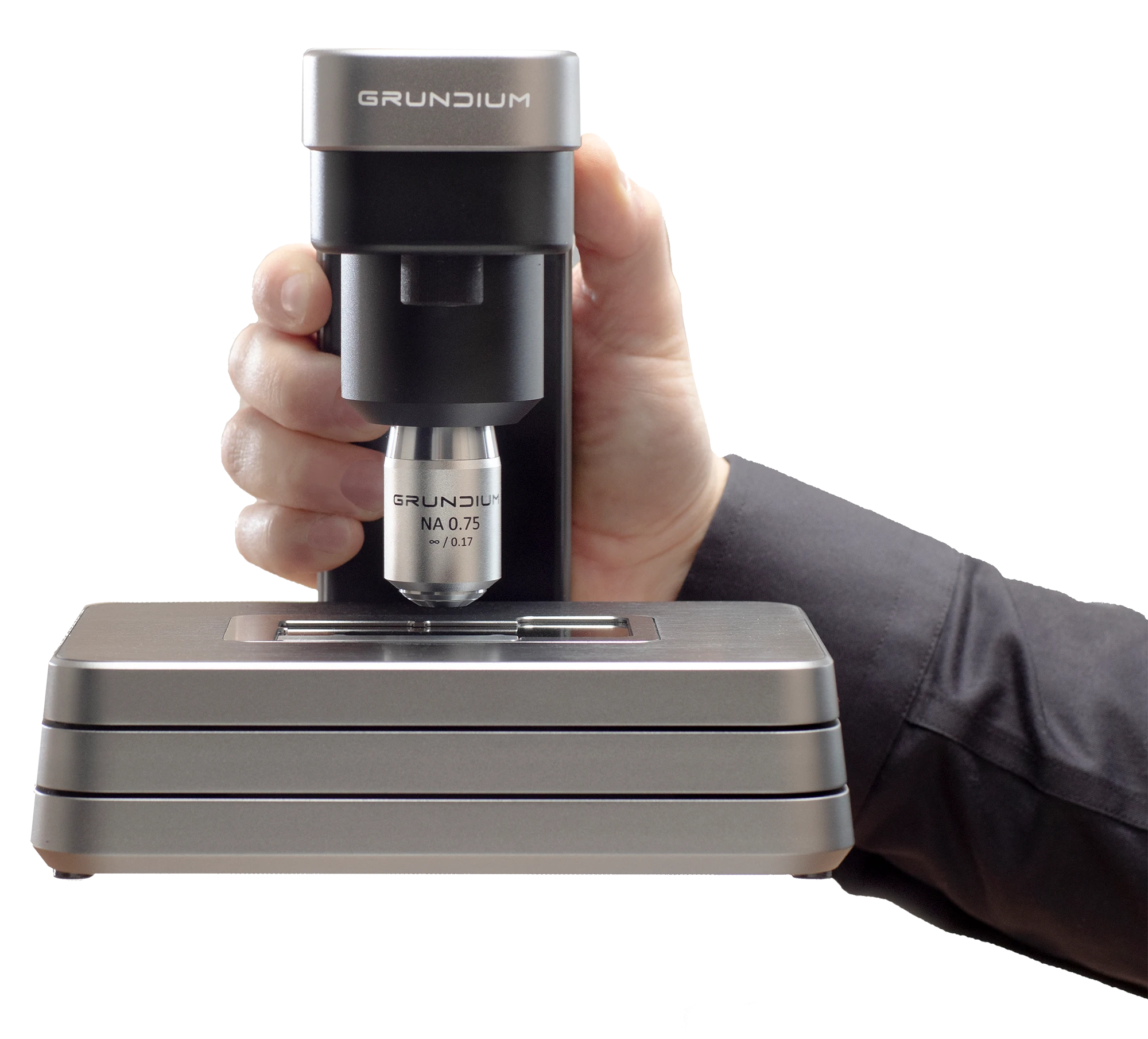
Ocus40 held in hand

4. WHAT KIND OF HARDWARE AND SOFTWARE REQUIREMENTS ARE NECESSARY FOR IMPLEMENTING WSI IN A PATHOLOGY LAB?

Hardware and software requirements depend primarily on the chosen scanner and the workflow in use. However, some common elements need to be considered in all whole slide imaging setups.

Slide Scanners: These are specialized devices that capture high-resolution images of pathology slides. They come in a range of sizes and capacities, from single-slide scanners to high-throughput devices capable of scanning hundreds of slides at a time.

Storage Solution: WSI produces massive data, requiring a substantial storage solution. This could be local servers or cloud-based storage. The choice depends on the lab's needs and resources. However, the solution must be robust, secure, and capable of handling large data transfers quickly.

Monitors: High-resolution monitors are recommended to display detailed WSI images effectively. Monitors used for digital pathology should ideally have a resolution of at least 2K (2560 x 1440 pixels), although 4K monitors are becoming increasingly common.
Network Infrastructure: Fast, reliable network connections are crucial for transferring and accessing large image files. Labs must have a robust network infrastructure with high-speed broadband or fiber-optic internet connections.

Integration Capabilities: To improve your workflow, consider how the different devices and software can be integrated to provide the most straightforward and smoothest setup for digital pathology.

Finally, successful implementation also requires consideration of factors like user training, system maintenance, and data security. It's also essential to have a disaster recovery plan in place to protect against data loss. Implementing a WSI system is a significant undertaking that requires careful planning and investment, but with the right resources and support, it can significantly enhance the practice of pathology.

5. HOW IS THE DIGITAL DATA FROM WSI STORED AND MANAGED, AND WHAT ARE THE IMPLICATIONS FOR DATA SECURITY?

The digital data generated by Whole Slide Imaging systems is often large in volume due to the high-resolution images they create. Storing and managing this data requires robust solutions that ensure accessibility, speed, and security.

Storage can be managed locally on servers within a healthcare facility or remotely through cloud-based solutions. Local storage often requires substantial investment in hardware and ongoing maintenance but provides direct control over data. Conversely, while requiring ongoing service fees, cloud storage offers scalability and accessibility from different locations, an advantage in telepathology, and for institutions with multiple sites.

The data management aspect involves structuring and organizing the data for easy retrieval, which can be facilitated through integration with Laboratory Information Systems (LIS). This integration also aids in annotating and linking WSI data with relevant patient and case information.
Healthcare institutions must adhere to stringent regulations regarding data security to protect patient confidentiality. These regulations, like the General Data Protection Regulation (GDPR) in the European Union, mandate the implementation of physical, network, and process security measures. This may include data encryption, secure data transfer protocols, access controls, and regular audits to ensure the integrity and confidentiality of patient data.

In summary, the handling of WSI data requires balancing the need for accessibility and usability against the need for robust data security measures to ensure that the digital pathology environment is both efficient and safe.

6. CAN WSI BE INTEGRATED WITH OTHER DIGITAL SOLUTIONS, SUCH AS ELECTRONIC HEALTH RECORD (EHR) SYSTEMS AND LABORATORY INFORMATION SYSTEMS (LIS)?

WSI systems can indeed be integrated with other digital solutions like Electronic Health Record (EHR) systems or Laboratory Information Systems (LIS). This integration can improve efficiency, allow for more streamlined workflows, and enhance patient care.

Integration with EHR systems allows seamless access to a patient's digital pathology slides alongside their other medical records, providing a holistic view of their health history. It can also facilitate better communication between pathologists and other healthcare providers, as the digital slides can be easily shared and discussed.

On the other hand, integration with an LIS can improve laboratory workflows. When WSI and LIS are integrated, the pathologist can often view the slide images and related metadata directly in the LIS. This reduces the need for switching between systems, improving efficiency and reducing the chances of errors.

However, successful integration does require careful planning and implementation, considering factors such as data security, system compatibility, and compliance with healthcare regulations. But when done well, the integration of WSI with other digital solutions can significantly enhance the practice of pathology.

7. WHAT TRAINING IS REQUIRED TO INTERPRET WHOLE SLIDE IMAGES EFFECTIVELY, AND ARE SUFFICIENT RESOURCES AVAILABLE FOR THIS?

Adopting Whole Slide Imaging in pathology practice requires some training, although the exact nature and duration can vary based on the individual's prior experience with digital tools and the specific WSI system in use.

Training typically involves understanding how to use the WSI software, including navigating and manipulating digital slides, adding annotations, and adjusting image settings..

Beyond these technical aspects, pathologists also need to learn how to interpret digital images effectively. While the principles are similar to those of traditional microscopy, the digital format can present unique challenges and opportunities. For example, the ability to zoom in and out quickly can change how pathologists approach slide reviews.
Fortunately, many resources are available for training in digital pathology. WSI system vendors often provide training as part of the installation process, and numerous online courses, webinars, and tutorials are also available. Several professional organizations, such as the Digital Pathology Association, offer resources and guidelines to support the adoption of digital pathology. Furthermore, conferences and workshops often have sessions dedicated to digital pathology, providing opportunities for continuing education.

So while a learning curve is involved in the adoption of WSI, sufficient resources are available to support pathologists in this transition. As with any new technology, the key to effective use is ongoing practice and continuous learning.

8. CAN WSI BE USED FOR ALL TYPES OF SPECIMENS, OR ARE THERE CERTAIN LIMITATIONS?

Whole Slide Imaging can be used effectively for a broad range of specimen types, including routine histological samples, cytology slides, and hematological smears. However, there are certain limitations to be aware of.

The quality of a digital image in WSI is reliant on the preparation of the physical slide. Some specimens, particularly those very thick or with high levels of three-dimensionality, can pose challenges for WSI. The focus of WSI scanners is usually set at a single plane, and specimens that exceed the focus depth may not be accurately represented in the digital image. However, this is not the case with Grundium’s Ocus scanners. They use an algorithm to select the best focus for each field of view, which makes the Ocus scanners excel in scanning uneven samples. After stitching the images together, the end result is a beautiful, sharp image without scanning artifacts.
Lastly, while advances have been made in scanning speed, WSI can still be time-consuming for very large specimens or those requiring high-resolution imaging, which could limit its use in urgent cases.

Despite these limitations, WSI technology is continually improving and proving increasingly valuable in pathology. Its capacity to create a permanent digital record of a slide and facilitate remote consulting, education, and integrated diagnostics are all substantial benefits that make WSI an attractive tool in modern pathology.

9. HOW MIGHT AI AND MACHINE LEARNING TECHNOLOGIES INTERACT WITH WSI TO ASSIST IN DIAGNOSES, AND WHAT ARE THE POTENTIAL IMPLICATIONS FOR PATHOLOGISTS' ROLES?

Artificial intelligence (AI) and machine learning (ML) technologies are becoming increasingly influential in the field of digital pathology, including Whole Slide Imaging.

These technologies can analyze digital images and identify patterns and features that may not be readily apparent to the human eye. They can assist pathologists by providing decision support, such as identifying areas of interest in a slide or classifying lesions. AI and ML could also help in quantifying features, such as counting cells in a smear, which could otherwise be time-consuming.

Importantly, AI and ML tools can increase efficiency by pre-screening slides and highlighting those with potential abnormalities, reducing the workload of routine cases and allowing pathologists to focus on more complex or urgent cases.

These technologies also hold promise in prognostic evaluations and personalized medicine by identifying subtle morphologic features that correlate with clinical outcomes or treatment responses.

However, while AI and ML have the potential to enhance pathology greatly, they do not replace the need for trained pathologists. The role of the pathologist may evolve, becoming more of a "diagnostic manager," integrating and interpreting information from various sources, including AI/ML algorithms.

It's also important to note that AI and ML models are tools that require oversight. They should be validated, and their performance should be monitored to ensure they're providing accurate and reliable results. As such, pathologists may also have a role in this validation and monitoring process.

Send us a message!

Do you have a question about our devices? Wondering how the Ocus® scanners could be implemented in your workflow? Whatever it is, we are happy to hear from you!

Send us a message with the form here, and our experts will get back to you shortly!

If you need assistance with your device, please contact Grundium's customer support.

About Grundium

Grundium is a global leader in digital imaging for pathology, transforming how medical professionals diagnose and treat diseases. Founded in 2015 and based in Tampere, Finland, the company leverages over two decades of expertise in optics and imaging to develop the Ocus® series of microscope scanners. These compact, high-precision devices make high-quality digital and remote pathology accessible to all, offering ease of use and affordability for labs of any size.

Trusted by healthcare providers worldwide, Grundium’s solutions enhance diagnostic accuracy, streamline workflows, and reduce unnecessary logistical steps—ultimately driving better patient outcomes through innovative technology.