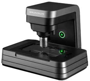
Whole Slide Imaging (WSI) refers to the process of scanning traditional glass pathology slides to create high-resolution digital images. These digital images, also known as "virtual slides," can be stored, shared, and analyzed using computer software, greatly enhancing the workflow in pathology labs. WSI enables pathologists to review slides remotely, collaborate with colleagues, and apply advanced computational tools, such as artificial intelligence (AI), for image analysis.
For example, WSI is often used in telepathology, where specialists can diagnose patients from afar. The technology is transforming the field of digital pathology, as it supports more efficient workflows, enhanced diagnostic accuracy, and better integration into electronic medical records.
Read our blog: 9 questions and answers about Whole Slide Imaging
Telepathology refers to the remote practice of pathology, where pathologists diagnose or consult on cases using digital images of slides transmitted over the Internet. This approach allows for real-time consultations or second opinions, even when the pathologist is not physically present at the lab.
Telepathology is particularly valuable for rural or underserved areas where access to specialized pathology expertise is limited. It is also widely used in research collaborations and educational settings. By leveraging telepathology, healthcare providers can offer faster and more accurate diagnostic services, reduce travel times and costs, and provide access to specialized expertise across various healthcare facilities.
A digital pathology scanner is a specialized device used to scan glass slides containing tissue samples and convert them into high-resolution digital images. These scanners enable pathologists to digitize traditional slides for remote viewing, analysis, and archiving. Most scanners offer magnifications ranging from 20x to 40x, allowing detailed examination of tissues and cellular structures.
Scanners vary in capacity; some handle a single slide at a time, while high-throughput scanners can process hundreds of slides simultaneously. This scalability makes digital pathology scanners ideal for both small diagnostic labs and large research institutions. High-resolution scanning plays a crucial role in telepathology, allowing pathologists to diagnose cases remotely or collaborate across geographic boundaries. Some scanners integrate with software systems to enhance workflows by automating slide labeling, storage, and image analysis.
A virtual slide is a high-resolution digital image of a pathology slide created using a digital pathology scanner. Virtual slides replicate the experience of viewing traditional glass slides under a microscope but offer numerous advantages. These include the ability to zoom in on areas of interest, annotate regions for future reference, and share the slide with colleagues instantly for consultations or education.
Virtual slides are essential in telepathology, where digital slides are used for remote diagnostics, allowing pathologists to provide expert opinions without being physically present. Virtual slides are also extensively used in pathology education, enabling students and professionals to view rare or complex cases without needing physical access to the original specimen. As a result, virtual slides significantly improve accessibility and collaboration in modern pathology.
A pathologist workstation is a dedicated computer system equipped with specialized software and hardware for viewing and analyzing digital pathology slides. With Grundium's Ocus® scanners, no specialized software is needed to view and annotate the images, as the user interface is browser-based. Typically, the workstation includes a high-resolution monitor for clear visualization of slides.
These workstations are designed to integrate seamlessly into a digital pathology workflow, allowing pathologists to quickly review cases, make annotations, and share findings with colleagues. In the future, more systems will also incorporate AI-based image analysis tools, helping to enhance diagnostic accuracy and streamline workflows. As digital pathology becomes more common, pathologist workstations are central to enabling efficient and accurate diagnoses in clinical settings.
Optical magnification refers to the degree to which a digital pathology scanner enlarges the image of a tissue sample using optical lenses. In digital pathology, scanners typically offer magnifications ranging from 20x to 40x, allowing detailed inspection of tissue architecture and cellular structures.
Higher magnifications provide more detail, making it possible to diagnose subtle pathological changes that might be missed at lower magnifications. This feature is critical in cases such as cancer diagnosis, where early detection of abnormal cells can lead to better outcomes. Digital scanners with adjustable magnification offer flexibility for pathologists, allowing them to switch between different levels of detail depending on the diagnostic requirements.
Read our blog: Magnification: Does it matter what is says on the tin?
Scanning resolution in digital pathology refers to the level of detail captured by a digital pathology scanner, typically measured in microns per pixel. Higher scanning resolutions allow pathologists to observe finer details within tissue samples, which is crucial for diagnosing certain diseases, especially those that involve small cellular changes.
Most scanners provide resolutions equivalent to 20x or 40x optical magnification, with higher resolutions offering better clarity for examining subtle tissue abnormalities. High-resolution scanning is critical in areas like oncology, where early detection of tumor cells can significantly impact treatment outcomes. For accurate diagnostics, laboratories often choose scanners that balance high resolution with fast scanning speeds to ensure both quality and efficiency.
Dive deeper: Resolution and scanning speed in high-power digital microscopy
In digital pathology, commonly used image formats include SVS (Aperio's proprietary format), TIFF (Tagged Image File Format), and DICOM (Digital Imaging and Communications in Medicine). Each format serves different needs in terms of image quality, file size, and compatibility.
SVS and TIFF formats are widely used for storing high-resolution pathology images with minimal loss of quality, making them ideal for detailed analysis. DICOM is a standardized format across medical imaging disciplines, ensuring interoperability between pathology software and hospital systems. The choice of image format impacts not only storage and performance but also the ability to share data efficiently with other healthcare providers and institutions.
A digital workflow in pathology refers to the end-to-end process of managing pathology cases digitally. This includes the scanning of glass slides, storing them in a digital format, analyzing images using advanced software, and generating reports for clinical diagnosis.
The adoption of digital workflows improves efficiency by reducing the manual handling of slides and enabling faster case reviews. Automated image analysis tools can assist in quantifying data, such as tumor size or cell counts, which enhances the accuracy and speed of diagnosis. Additionally, digital workflows facilitate collaboration between pathologists and clinicians across different locations, making it easier to share cases and offer consultations.
An Image Management System (IMS) is software designed to organize, store, and manage digital pathology images and related data. These systems help pathologists efficiently retrieve, review, and archive digital slides, ensuring that each image is associated with the correct case.
An IMS typically integrates with hospital information systems (HIS) and laboratory information management systems (LIMS), allowing for seamless case tracking, diagnostic workflows, and reporting. Many image management systems also offer features like real-time collaboration, access control, and automated notifications, which help streamline pathology workflows. By optimizing the storage and retrieval of large image files, an IMS enhances productivity in both clinical and research environments.
Artificial Intelligence (AI) is revolutionizing the field of digital pathology by enhancing diagnostic accuracy, speed, and consistency. AI in pathology typically involves the use of machine learning algorithms and deep learning models trained to recognize specific patterns, abnormalities, or structures in digital images. These AI models can assist pathologists by automating routine tasks like cell counting, tumor detection, and tissue segmentation, allowing for faster case reviews.
AI's potential extends to areas like cancer detection, where it helps identify malignant cells at early stages and predict disease outcomes by analyzing patterns that may not be immediately obvious to the human eye. Moreover, AI tools are often designed to integrate seamlessly into digital workflows, ensuring that they support, rather than replace, pathologists in making more informed decisions. As AI continues to evolve, it will likely play an increasingly important role in reducing diagnostic errors and improving patient outcomes.
One of the key benefits of AI in pathology is its ability to handle large-scale image data, making it an essential tool for high-volume labs. However, the adoption of AI comes with challenges, such as regulatory approvals, data privacy, and the need for well-curated datasets to train the models effectively.
Our articles about AI:
Zoetis & Grundium: An example of telepathology & Artificial Intelligence in remote imaging at satellite clinics
Artificial Intelligence vs. Human Expertise in Pathology
Immunohistochemistry (IHC) is a critical technique in pathology that allows for the visualization of specific antigens in tissue sections through the use of labeled antibodies. In the context of digital pathology, IHC is increasingly being digitized to enhance precision and reproducibility in diagnostics. Once stained, IHC slides can be scanned by digital pathology scanners and then analyzed using software tools designed to quantify the presence, intensity, and distribution of biomarkers within the tissue sample.
Digital IHC is especially valuable in cancer diagnosis and personalized medicine, where identifying specific proteins or gene expressions can help determine the type of cancer and guide treatment decisions. For example, in breast cancer pathology, IHC is used to assess the expression of hormone receptors and HER2 status, which are crucial for determining therapy options.
Regulatory compliance is a vital aspect of implementing digital pathology in clinical settings. In the United States, the Food and Drug Administration (FDA) regulates the use of digital pathology systems for primary diagnosis, ensuring that they meet stringent standards for accuracy, safety, and reliability. Similarly, other regions, such as the European Union, follow regulations like the In Vitro Diagnostic Regulation (IVDR) to ensure that digital pathology tools comply with medical device standards.
Compliance typically involves a thorough validation process where digital pathology systems, including scanners, software, and AI tools, are rigorously tested to ensure they produce diagnostic results that are as reliable as those obtained through traditional methods. This includes evaluating the image quality, scanner accuracy, and any software used for image analysis.
One key aspect of regulatory compliance is ensuring that digital pathology systems are FDA-approved or CE-marked (for the EU market) before being used for clinical diagnoses. Moreover, labs must adhere to standards for data security and patient privacy, especially when using cloud-based storage solutions.
Barcode tracking in pathology refers to the use of barcodes to track tissue samples, slides, and corresponding data throughout the pathology workflow. In a digital pathology setting, barcode tracking ensures that each sample is accurately labeled and traced from the moment of biopsy through to diagnosis and archiving.
Barcodes are essential for maintaining the integrity of the sample identification process, reducing errors in labeling, and ensuring that the correct diagnosis is associated with the correct patient. A typical barcode system is integrated with Image Management Systems (IMS) and Laboratory Information Management Systems (LIMS), which help automate sample tracking, improve turnaround times, and enhance workflow efficiency.
Barcode tracking is vital in high-volume labs where many samples are processed simultaneously. By automating sample identification and minimizing the risk of human error, barcode tracking systems improve both accuracy and productivity. Moreover, these systems ensure that the chain of custody is maintained, making them invaluable in research settings and clinical trials where data integrity is paramount.


