


Out of all the scanners I’ve used — and I’ve used a lot — it’s the easiest one. We've scanned more than 30 000 slides without any technical issues.

With Grundium Ocus-enabled telepathology, we can provide better service to our customers; the hospitals. For the hospitals the process doesn’t change, but they benefit from Fimlab working faster and more accurately with digital pathology.

Not only has Grundium created something unique in the market with the beautifully designed small-footprint scanner, but they also have a super technical team that can work really close together to make the whole solution seamless and is willing to invest themselves in making something new. It’s super unique in the world to have everything from the region of interest, focus layers, depth of field and the whole experience totally integrated.

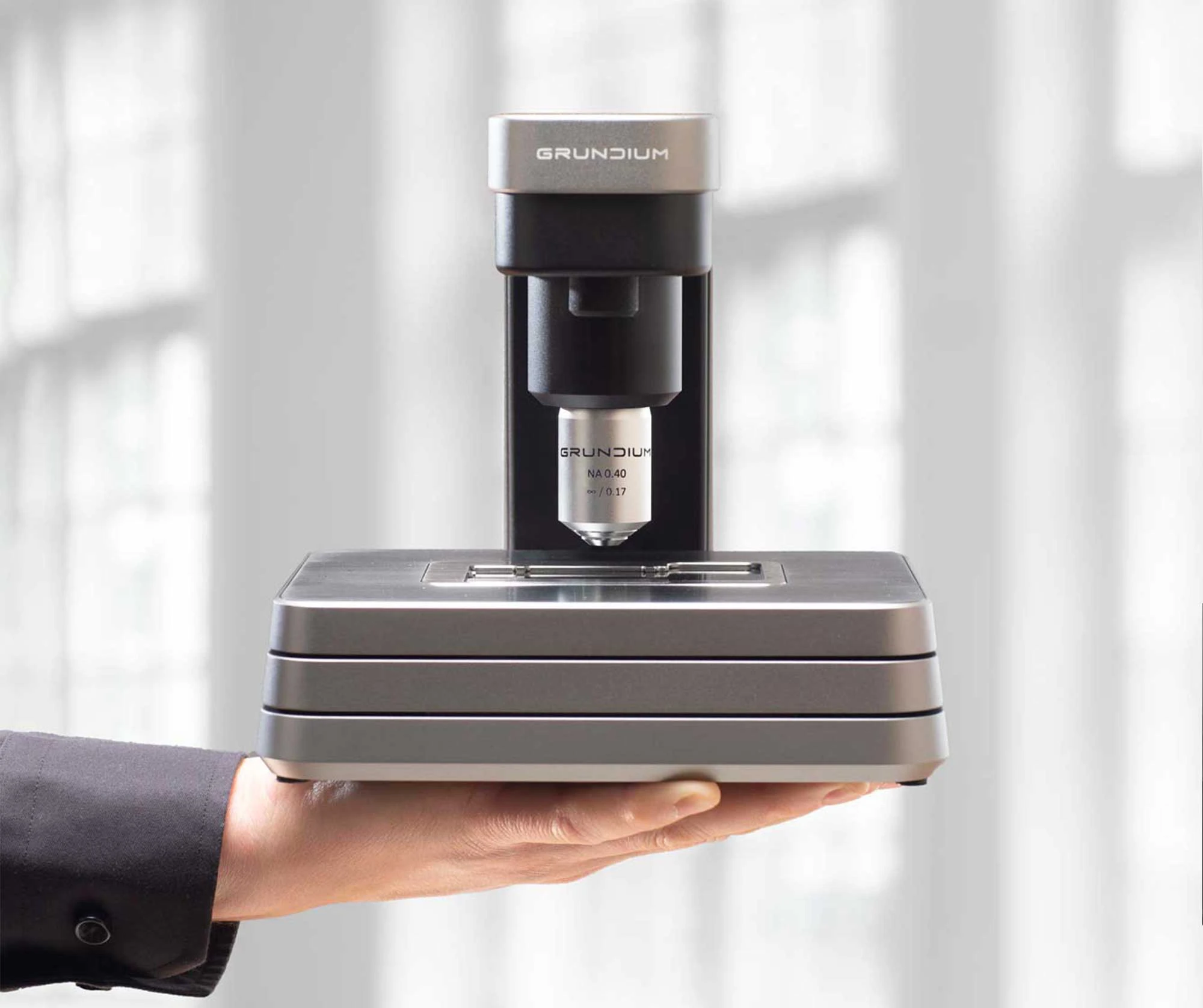
The Ocus scanner is small and produces very sharp images fast. It is a vital instrument in field use in organ procurement. Its unparalleled portability helps save lives.

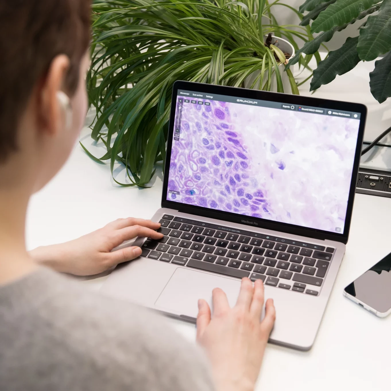
We have excellent images with the Ocus and I couldn’t be happier to have this little helper on my desk here. I use it mainly as a microscope, to be honest, but of course, scan my slides as well. It has really helped me so much.

The Philips IntelliSite Digital Pathology Solution has the capabilities to upload, view and annotate the images produced from the Grundium scanner. Zooming and panning is really fast, even when the images are physically stored on a server in the Netherlands and I’m viewing the images from a workstation in Sweden.
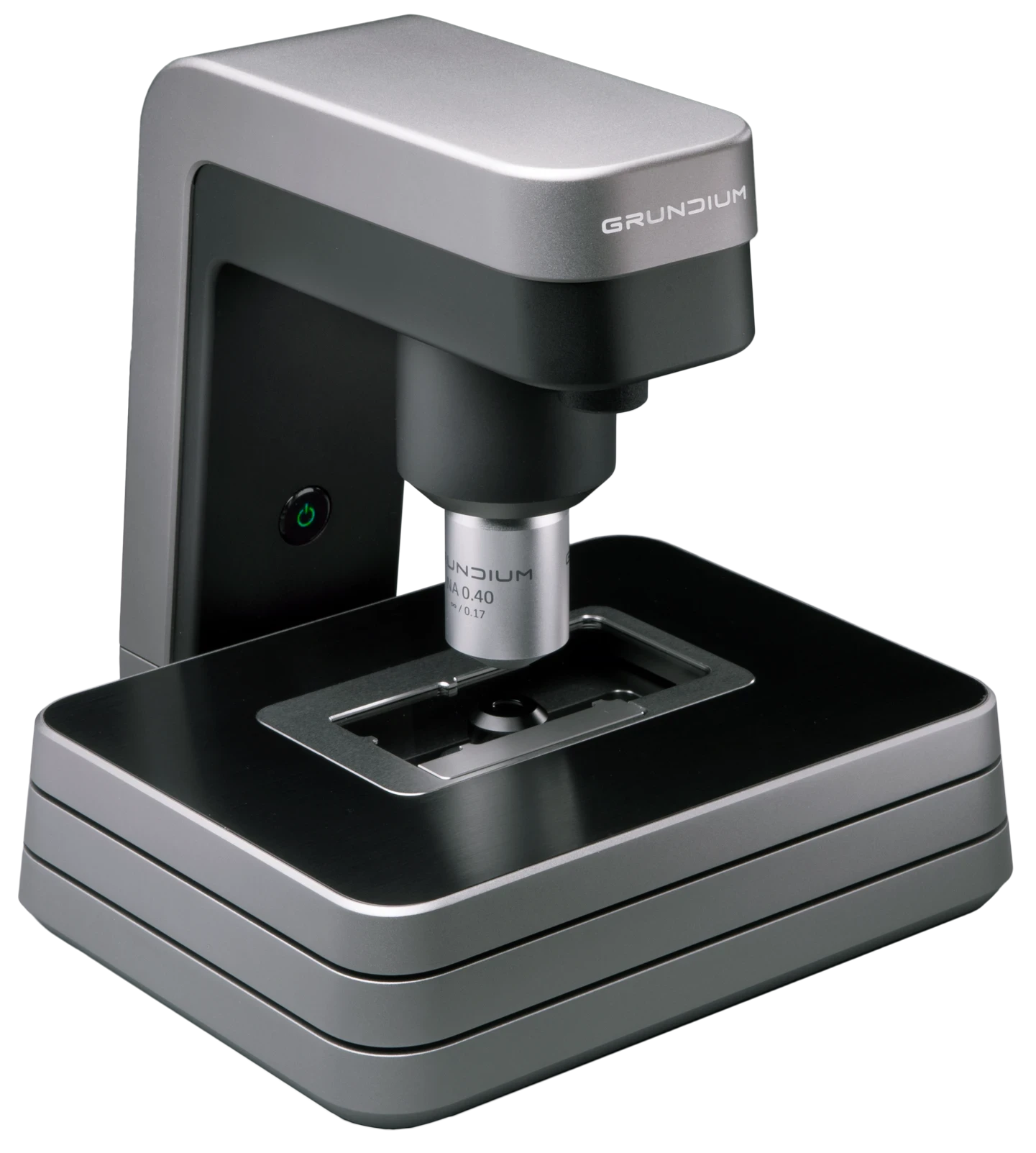
Magnification: 20x
Numerical Aperture: 0.40
Resolution: 0.50 µm / pixel
Depth of field: 5 µm
Slide format: 75 x 25 mm (3 × 1 in)
Scan speed: ~60 sec / 15 × 15 mm
Image formats: .SVS, .TIFF, .SZI
Focusing: Fully automatic
Image sensor: 12 MPix
Dimensions: 7 × 7 × 7.5 inches (18 × 18 x 19 cm)
Weight: 7.7 Lbs / 3.5 Kg
Internal Storage: 500 Gigabytes
Power consumption / Standby: 0,03 A, 0,007 kVA, 0,007 kW
Power consumption: / Operation: 0,09 A, 0,021 kVA, 0,021 kW
Grundium Ocus is a digital microscope slide scanner designed to convert glass slides into high resolution digital images for use in digital pathology workflows. As a 20x slide scanner, it belongs to the category of digital microscope slide scanners and whole slide scanners. Producing digital slides and whole slide images from standard microscope slides for diagnostic and research use.
Ocus®20 is used for slide scanning in routine digital pathology workflows, including intraoperative frozen sections, remote consultations, and research use. It supports automatic scanning of slides and helps improve workflow efficiency by enabling reliable digitization of pathology slides for review and analysis.
A 20x pathology scanner is commonly used because it offers a practical balance between diagnostic detail and workflow efficiency. For many routine histology applications, 20x provides enough resolution to assess tissue architecture and most key morphologic features, while keeping scanning times predictable.
Common 20x use cases include routine surgical pathology on H&E slides, many biopsy workflows, and a large portion of immunohistochemistry review and scoring. It is also widely used for consultations, tumor boards, teaching, and digital archiving where speed and file manageability matter.
Ocus®20 is designed to scan standard glass slides and microscope slides used in pathology laboratories. It supports common sample types encountered in histology and frozen section workflows, converting physical slides into digital slides suitable for detailed analysis.
Ocus®20 supports workflow efficiency through automated scanning, fast scan times, and consistent image quality designed to minimize rescan rates. This helps laboratories handle routine workloads smoothly, while also supporting time-sensitive cases by keeping scanning predictable and reducing interruptions.